AI in medical imaging: from research object to omnipresent helper
The practical use of artificial intelligence in medical imaging is progressing: what was previously mainly the subject of scientific research is now finding its way into daily routine - in hospitals, but also increasingly among practising radiologists. The demand is enormous.
Life expectancy is increasing worldwide: according to the WHO, the number of people over 60 is set to double to 2.1 billion by 2050. As a result, the need for care in the treatment of diseases such as cancer and osteoporosis is growing, as is the demand for medical imaging. Computed tomography (CT), magnetic resonance imaging (MRI), ultrasound and good old-fashioned X-rays are all indispensable diagnostic tools.
According to statistics, 125 million X-ray examinations and around 13.5 million CT and MRI scans were carried out in Germany in 2021. However, according to the OECD, there are far too few specialists to analyse all the data produced. In addition, errors can occur under time pressure - the number of errors worldwide is estimated at several million per year.
Fewer errors, more accurate diagnoses
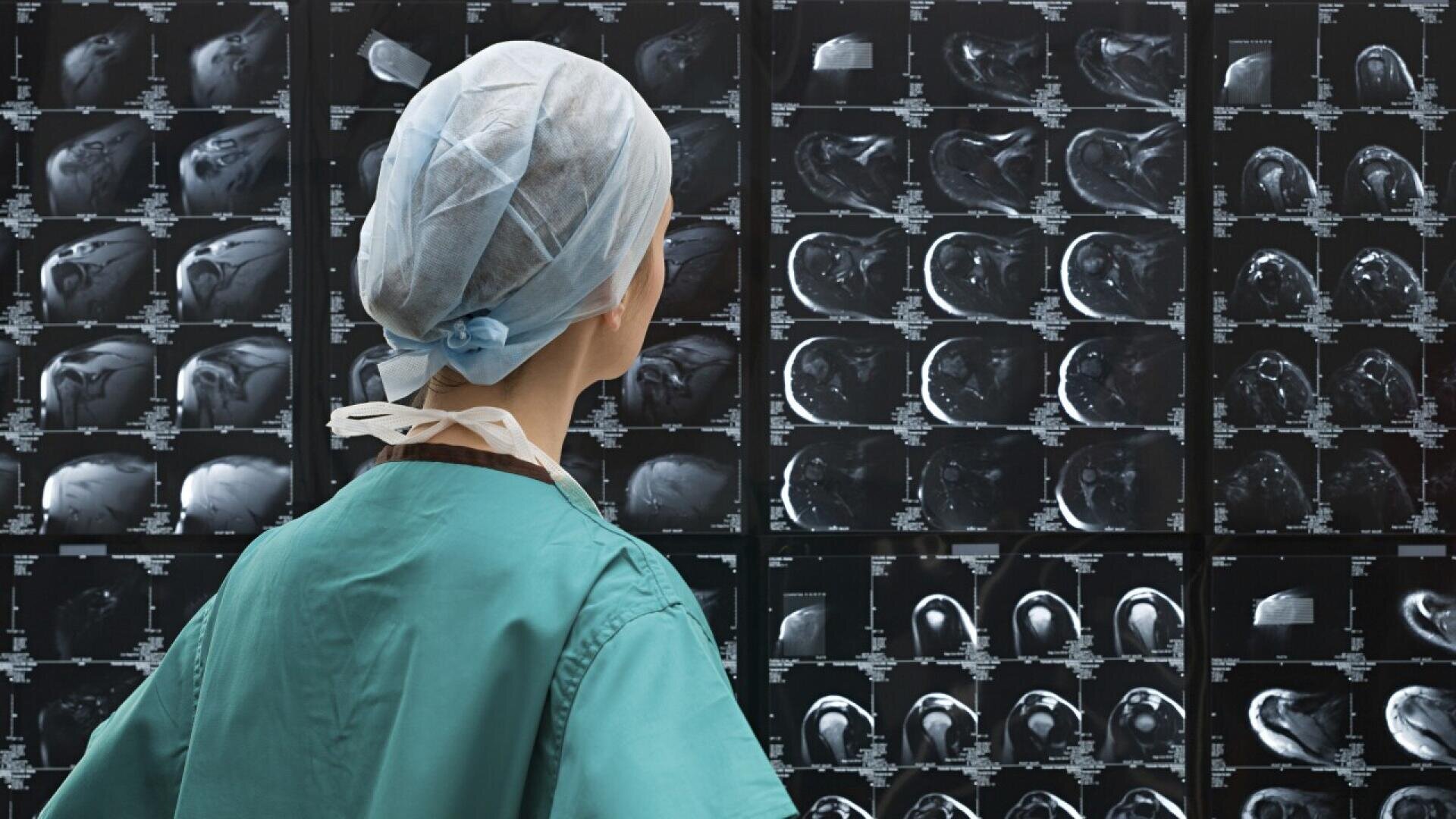
Artificial intelligence can provide valuable services when trying to relieve the burden on medical staff: ‘Most medical images are extremely data-intensive and it is very time-consuming to manually check all areas of an image. It is also easy for a human to overlook information in an image. AI helps to reduce the analysis time and helps the doctor to focus on the most important parts of an image,’ says Dr Katherine Fitch from the Fraunhofer Institute for Cognitive Systems IKS in Munich. Together with her team, the head of the Trustworthy Digital Health department is developing explainable, robust and unbiased AI models for the healthcare sector.
But it's not just about avoiding errors. It is undisputed that AI can raise the quality of examination results to a new level. Katherine Fitch: ‘AI can achieve greater accuracy in tasks that involve characterising an image, for example when it comes to the question of how large the left atrium of the heart is. This is very important for more accurate diagnoses and treatment decisions.’

Katherine Fitch, Fraunhofer IKS © Fraunhofer IKS
Large number of projects
Favoured by the exponentially increasing computing power of computers, researchers like Fitch are working in many places to find out how AI can be used for imaging. Advanced algorithms and neural networks, developed and trained according to deep learning methods and using a large amount of medical image data, analyse with an accuracy that surpasses the human eye. There are now a large number of projects in the clinical field that have raised great hopes.
To name a few examples:
- In the MIDAS project, the University of Tübingen is researching imaging applications and computer technologies that enable reliable, explainable solutions that can be interpreted by humans for the integration of AI into clinical practice and offer patient-centred workflows for the diagnosis and treatment of neurological, cardiovascular and oncological patients.
- In 2024, a collaboration between Ludwig-Maximilians-Universität München, TU Berlin and Charité resulted in a new AI tool that can also recognise less common diseases in the gastrointestinal tract based on imaging data without having to be specifically trained for these rarer cases.
- Last year, researchers at TU Graz succeeded in generating precise real-time images of the beating heart from just a few MRI measurement data with the help of cleverly trained neural networks. This method could make MRI applications faster and therefore cheaper in the future.
Nevertheless, progress with AI in everyday clinical practice in hospitals and surgeries has been comparatively slow to date, not least due to the availability of patient data for training purposes. As this data is highly sensitive and particularly worthy of protection, access to it is restricted and only possible under strict conditions.
AI on its way into practices
However, what has long been limited to research projects is now well on the way to being widely used in the healthcare sector and therefore also in the approximately 2,000 radiological practices in Germany. An important prerequisite for this is that the findings generated with the help of AI are also rewarded accordingly. There are two options for this: the statutory health insurance funds reimburse the additional service, or it is offered to patients as an ‘individual health service’ (IGeL).
The first option now appears to be in place: It was only at the end of November 2024 that IKK Südwest announced that it was setting a ‘milestone’ in the integration of AI technology into healthcare systems. It concluded a reimbursement agreement with the start-up Contextflow. The Vienna-based company has developed AI-based software that should enable radiologists to detect lung cancer up to one year earlier and with significantly greater accuracy. This could help to reduce healthcare costs.
Fully automated assessment of bone health
Osteoporosis is another widespread disease that causes high costs in the healthcare system due to the existing diagnostic gap. One in three women and one in five men will suffer a sometimes life-threatening osteoporotic fracture in the course of their lives. More than two thirds of patients who should be treated according to the current osteoporosis guidelines do not receive the appropriate treatment in Germany.
Bonescreen addresses this problem. The Munich-based start-up has developed an AI-based technology to support radiologists and clinicians in assessing bone health. ‘Our product uses artificial intelligence to use existing CT scans, which are performed in routine clinical practice anyway, fully automatically to assess bone health and thus extract information in a resource-saving manner without changing the infrastructure or workflows in order to prevent bone loss in the population,’ explains Dr Sebastian Rühling, co-founder and Chief Medical Officer of Bonescreen.

Sebastian Rühling, Bonescreen © TU München
Evidence to convince health insurance companies
How his technology works: the bone density, which is actually invisible to the human eye in a CT scan, is quantified by the AI and also displayed in a coloured heat map. This visualisation makes even subtle differences visible, allowing radiologists to make an objective assessment of bone density. Existing bone fractures, some of which have gone unnoticed and silent, are recognised.
Sebastian Rühling expects CE certification in spring 2025. The procedure, which has mainly been used in clinical research groups to date, is then to be rolled out on the market - primarily in the practices of registered radiologists, initially as an IGeL, with the aim of being reimbursed by statutory health insurance funds in the medium term. Sebastian Rühling: ‘We believe that we can convince the stakeholders in the healthcare system of the evidence and thus enable reimbursement by the statutory health insurance funds. We want to prove that it is cheaper for the healthcare system to measure bone density for a few euros than to bear the follow-up costs. Our focus is on continuously increasing the benefits for users, patients and society. We have therefore placed particular emphasis on a broad database to ensure that the models not only work in a small specific collective, but also in hospitals and surgeries and that any scanner currently on the market can be used, for example.’
Contextflow and Bonescreen are not the only companies that are now launching. Other start-ups that have emerged from the university environment are preparing to conquer the radiology market.
Device manufacturers are breaking new ground
The manufacturers of imaging devices are already at home there. They are also focussing on artificial intelligence and equipping their scanners with it. ‘In the field of MR imaging, we have succeeded in speeding up examination times by almost 80 per cent using AI-supported image reconstruction. A knee examination that previously took 10 minutes on a 3 Tesla device can now be completed in two minutes. This is more comfortable for patients and also means that more examinations can be carried out in the existing time, reports Tobias Heimann, Head of Artificial Intelligence Germany at Siemens Healthineers.
Another path that hardware manufacturers are pursuing is the progressive miniaturisation of their devices. Healthineers is also making its contribution here: ‘Let's take MRI as an example. Until now, a good half of humanity has not had access to this technology. We have succeeded in developing a device that is smaller, more compact and independent of the rare resource helium. This means that it can also be used in places where this was previously not the case, for example because there was no liquid helium for magnetic cooling or because previous devices were simply too large and too heavy for installation,’ says Tobias Heimann. Another example of the trend towards miniaturisation is a CT device that has been developed for use in special ambulances, so-called mobile stroke units. Tobias Heimann: ‘It enables a precise diagnosis to be made in the ambulance, in a quality that we are otherwise only familiar with from stationary devices. This means that patients can receive the right treatment more quickly and no valuable time is lost, which is crucial in the event of a stroke.’

Tobias Heimann, Siemens Healthineers © TU München
Language models optimise the workflow
Improving workflows in hospitals and surgeries is another goal of the use of artificial intelligence that could become very important in the coming years. If AI supports the optimised use of scarce resources, acceptance among medical staff can be further increased, as Dr Sebastian Bickelhaupt, radiologist and Clinician Scientist at the Institute of Radiology at the University Hospital Erlangen, explains: ‘Among the broad spectrum of possible applications for artificial intelligence in radiological imaging, we see great potential in supporting radiological workflows in hospitals and practices. This can include, for example, AI-based acceleration of image acquisition, support in the execution of examinations and the pre-stratification and automatic quality control of image data.’
Further developments of large language models (LLM) could also play a role in radiology, says Sebastian Bickelhaupt, ‘for example, to structure findings and automatically compile important information for reporting - provided that the regulatory requirements for data protection and data security in medical care, among other things, are met.’

Dr. Sebastian Bickelhaupt , Radiologischen Institut am Universitätsklinikum Erlangen ©Dr. Sebastian Bickelhaupt
Further applications for generative AI
Generative AI is apparently also being considered at Healthineers. The company sees data preparation, scan optimisation, diagnostic support and improved patient communication as potential areas of application for large language models and the technology on which they are based, the so-called ‘foundation models’. Tobias Heimann says: ‘AI solutions currently available on the market are generally focussed on clearly defined tasks, such as the automatic recognition of a specific anatomical structure or pathology and were also created specifically for this purpose. This requires a large amount of training data that has to be carefully prepared by hand. Foundation models can be trained without these time-consuming manual annotations and then serve as the basis for a large number of different tasks. This is one of the major technological trends, especially when multi-modal data such as images and text are used together.’
Katherine Fitch from Fraunhofer confirms that there will be interesting applications for generative AI in imaging: ‘Collecting enough data to train AI models is a problem today. This is where generative AI could really help. It can also be used to provide text-based comments on medical images.’ However, it will be important to make it as secure as possible: ‘In a sensitive application like healthcare, it is very important that we can understand the decision-making process of a model. Decisions should be made on the basis of important characteristics and not random correlations. Only then can a doctor effectively evaluate the AI recommendations to provide the best possible treatment for the patient,’ Katherine Fitch points out.
Advances in imaging technology, made possible by AI, are transforming medical diagnostics. These technologies offer the opportunity to make diagnoses more precise, faster and more cost-effective. At the same time, they reduce the workload of medical staff and improve patient care.
MedtecSUMMIT focuses on the use of AI
MedtecLIVE, the leading European trade fair for the development and manufacture of medical technology, gives companies along the entire value chain a platform to present their latest developments in this field to an interested trade audience. MedtecLIVE is part of a brand family that offers the industry more than just a trade fair and supports it in advancing medical technology with its wide range of products and services as well as its strong network and partners. This creates several platforms at the Stuttgart, Nuremberg and Munich locations, where industry players can meet their target group in a customised way.
‘In a constantly evolving world, it is crucial that we shed light on the latest advances. The next opportunity to showcase innovations, products and services, gain valuable impetus and initiate collaborations will be at our Innovation Expo trade exhibition as part of MedtecSUMMIT in Nuremberg from 18 to 19 February,’ says Silke Ludwig, Deputy Director MedtecLIVE. The summit is dedicated to the topics of clinical robotics, digital health applications, the utilisation of health data, innovations in sustainable materials and the use of intelligent implants. A separate thematic focus will address the question of how artificial intelligence can improve diagnosis, therapy and aftercare by analysing data. Experts from science, practice and industry will provide answers.

Silke Ludwig, MedtecLIVE © NürnbergMesse


